When you have a medical scan, it’s usually an MRI (magnetic resonance imaging), CT (computed tomography) or more recently, PL imaging (photoluminescence). Sometimes it’s all three as your care team works to determine what’s ailing you. That means three different appointments and three different imaging agents — typically nasty-tasting stuff you have to drink in order to sufficiently enhance the imaging signal so that diseased tissue can be distinguished from healthy tissue. Each comes with its own side effects and potential risks.
“What the medical field has long needed is a single imaging agent that will work across multiple imaging systems,” said Adah Almutairi, PhD, associate professor in the Skaggs School of Pharmacy and Pharmaceutical Sciences at UC San Diego.
Almutairi is always one to take up a challenge like that. Her bioresponsive materials lab is known for designing and developing smart polymers, nanoparticles and hydrogels for many innovative medical and research applications. One of Almutairi’s pet interests is in lanthanides, a family of naturally occurring chemicals that intrigued 19th century chemists because, among many other interesting properties, they burn easily in air, fluoresce under UV light and react with most nonmetals.
Inexplicably, scientific interest in lanthanides waned in the 1970s. A couple of years ago, Almutairi took up the mantle to explore how lanthanides do one special thing: convert low energy light into high energy light. She has long believed that her team could take advantage of that property for medical applications.
Almutairi and her team recently developed a new nanoparticle with a lanthanide-based core-shell-shell architecture. The nanoparticle emits light for optical imaging, but also relaxes water molecules for MRI and attenuates X-rays for CT simultaneously.
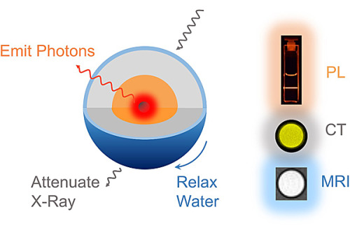
Three in one: the lanthanide nanoparticles can be used for photoluminescence (PL), computed tomography (CT) and magnetic resonance imaging (MRI) simultaneously.
In a study published in Nano Letters, the researchers tested these nanoparticles in “phantom” tissue — a hydrogel system that mimics living tissue in the laboratory. Not only does the nanoparticle work for each imaging type, it works better than each individual imaging agent on its own.
The team is now working to reduce the size of their new imaging nanoparticle so a patient’s kidneys can clear it more easily from the bloodstream.
“The main point of this study is that we overcame an engineering challenge,” said Sha He, a graduate student in the Jacobs School of Engineering at UC San Diego and first author of the new study. “Now we will tweak the design so we can advance this technology to pre-clinical and clinical testing. Our goal is that one day this nanoparticle, or one like it, will allow a patient to complete his or her imaging all at once, reducing the risk and toxicity associated with separate administration of multiple imaging agents.”
No comments:
Post a Comment