Showing posts with label nanoengineering. Show all posts

Thursday, March 31, 2022

Shaochen Chen receives NSF BRITE Award to develop 3D printed microscale human organs-on-a-chip

NSF Brite Fellow Shaochen Chen of the University of California San Diego will create a nanoscale biomanufacturing platform powered by artificial intelligence that can serve as a functional, three-dimensional model of human tissues, organs and systems. This “human-on-a-chip” could transform regenerative medicine and tools for disease modeling, drug screening, environmental studies, space exploration, and other applications. 

The National Science Foundation has awarded an engineering research grant to Shaochen Chen, professor and chair of nanoengineering at the University of California San Diego, to explore 3D printed microscale human organs-on-a-chip. 

Funded through the NSF Boosting Research Ideas for Transformative and Equitable Advances in Engineering (BRITE) program, the 5-year BRITE Fellow grants provide up to $1 million per project. The NSF BRITE program is funded by NSF Division of Civil, Mechanical and Manufacturing Innovation (CMMI) in the NSF Engineering Directorate. 

"NSF created the BRITE Fellows opportunity to support proven researchers who have a high-risk vision with the potential for high reward for the Nation," said CMMI Division Director Robert Stone. "We anticipate that the research of the BRITE Fellows will create new fields, challenge current paradigms, or present unconventional approaches to intractable problems."

With funding from the NSF Brite program, Chen will create a nanoscale biomanufacturing platform powered by artificial intelligence that can serve as a functional, three-dimensional model of human tissues, organs and systems. This "human-on-a-chip" could transform regenerative medicine and tools for disease modeling, drug discovery and screening, environmental toxicity studies, and other applications.

By powering his 3D bioprinting platform with artificial intelligence, Chen aims to minimize the trial-and-error associated with the process that often leads to waste of cells and time. "This is a critical issue for bioprinting since live cells are quite expensive, difficult to acquire (think of patient cells, for example), and do not survive well outside the incubator," he said. 

The ultimate goal of the project is to fabricate a human-on-a-chip in a microfluidic platform that integrates major human tissues including heart, liver, kidney, lung, and gut, and then study the chip's biomechanics and tissue functions. 

Friday, January 29, 2021

FAQs about UC San Diego’s wearable mask sensor for COVID-19

Monitoring your potential exposure to COVID-19 could be as simple as wearing a sticker on your mask. Researchers at UC San Diego are developing test strips that can be stuck on N95, surgical or cloth masks and be used to detect the presence of SARS-CoV-2 in a person’s breath or saliva. 

This newly funded project has made headlines—and raised questions. For answers, we followed up with Jesse Jokerst, a professor of nanoengineering at UC San Diego who is leading the project. 

How is this approach different from other at-home tests for COVID-19? 

This approach is designed for daily COVID-19 surveillance in high-density settings where a lot of people are together indoors for prolonged periods of time, such as hospitals, nursing homes, shelters, prisons, dialysis centers, halfway houses, etc. Our goal is to facilitate early detection of COVID-19 infections in high-risk populations. This approach provides a simple way to do that by integrating daily detection onto something that people are already wearing (masks). The notion is that a new day means a new strip; with this constant nature of surveillance, we can stop outbreaks early in high-density settings before they escalate. And rather than requiring you to swab your nose or throat, this strip collects particles that you breathe in and out throughout the day. 

How do the stickers work?

The stickers are designed to detect the presence of protein-cleaving molecules called proteases—not the virus itself—that are produced from infection with the SARS-CoV-2 virus.

The idea is that a sticker would be worn on your mask, in front of your mouth.


Throughout the day, airborne particles that you inhale and exhale will accumulate in the test strip.


When you’re ready to activate the strip, you would peel the sticker off and squeeze the blister pack.


The blister pack releases a reagent that changes color (for example, pink to purple as illustrated here) in the presence of the SARS-CoV-2 proteases. The top line of the strip is the control line, the bottom line is your test line. A color change in the reagent at the bottom line would indicate a potential exposure; this means you should get tested for COVID-19.


You would then dispose the used sticker and replace with a new one.

Can I use the stickers in place of getting a COVID-19 test?

The stickers are not a replacement for getting a COVID-19 test. They are not as sensitive as diagnostic COVID-19 tests run in a lab. The stickers are meant to be used as a surveillance tool for monitoring COVID-19 exposure, which can be done on a daily basis. Diagnostic testing, on the other hand, is done occasionally or less frequently. That’s the difference between testing and surveillance.

The analogy I like to use here is that this is like having a smoke detector in your home. It sits in the background at the ready and when it goes off, it does not tell you exactly where the fire is or what kind of fire it is, but that there is a problem and you need to take action. With these stickers, we’re similarly creating a warning device to tell you that you may have been exposed to SARS-CoV-2, and that’s when you would seek formal testing.

Why would we need these stickers if we already have COVID-19 tests?

While diagnostic viral testing is a critical tool, it does not scale readily for daily monitoring of asymptomatic or pre-symptomatic individuals. The value of surveillance for infectious disease is that it is ongoing. Just like how your smoke detector is in the background and at the ready, the stickers would be working on something you’re already wearing and can be used daily. This approach would supplement other monitoring efforts like wastewater monitoring, which is currently happening at UC San Diego. The stickers would add to our arsenal in the fight against COVID-19 and help us identify outbreaks early before they have a chance to spread.

Does a positive result mean I am infected with COVID-19?

Just like your neighbor’s house fire could cause your smoke alarm to go off, a positive result does not necessarily mean that you have COVID-19. It does mean potential exposure, so you should move beyond the surveillance phase and seek active testing. This could help identify asymptomatic and pre-symptomatic cases early, and thus prevent outbreaks in high density settings. 

How will the sticker detect what’s in my breath or saliva if it’s placed outside my mask?

Most masks have some airflow (otherwise you would suffocate). The sticker has vents both at the back—that allow air to flow through from your mask—and at the front to sample air from the environment.

If the sticker is meant to work outside the mask, does this mean that masks are not protecting us from COVID-19?

Masks definitely prevent the spread of COVID-19. All masks have some degree of permeability. N95 respirators, for example, reduce particle spread by 95%, so 5% of airborne particles can go through. This is the permeability we are exploiting to sample the air.

Will the sticker also detect SARS-CoV-2 particles in the air around me?

We do not know yet but hope so. The sticker will accumulate aerosols that you breathe in and out. The sticker will sample the air in your environment—not just the air that you are exhaling. Our goal is that this would serve as surveillance of the environment and not just the person wearing the sensor.

Can I wear the sticker inside my mask instead of outside?

Yes, either should work but we suspect that the exterior would be more comfortable. 

Would wearing this sticker be a violation of privacy? 

No. The sticker does not change color spontaneously as the user is wearing it. The color change would occur when the user activates the strip, which would be done in private.

How long would I need to wear the sticker before performing the test? 

We designed this for use in high density settings where individuals are at high risk, like nursing homes, shelters and prisons. The goal is that at the end of each 8-hour shift, residents, patients and staff would activate their test strips. We are doing studies to determine if shorter time periods can activate the color change. 

Can I still use my mask after activating the test strip? 

Absolutely. Just peel off the sticker when you are done. 

When will this be available to the public? 

The stickers are still in development. We currently have the reagents (in the blister pack) validated with recombinant proteases and are now evaluating them in a variety of more complicated samples. One of our next steps is to test the strips on COVID-19-positive saliva samples. We will work with UC San Diego School of Medicine Professors Louise Laurent and Rob Knight, who have access to a biobank of these samples. 

One big question is: How much of these proteases accumulate on our face coverings? And that depends on how long we wear them on our faces and how much we speak. Once we have these questions answered, we will begin testing in a cohort of known positive and known negative subjects. This should be in the second quarter of 2021. 

Then, we will move on to testing on patients and healthcare workers at VA San Diego Healthcare System, in collaboration with William Penny, a professor of clinical medicine at UC San Diego School of Medicine and a cardiologist at VA San Diego Healthcare System. 

Could this work as a saliva test? 

If these do not work out as mask stickers, we will repurpose the strips as dipstick tests using saliva as a sample. Here, the user would simply expectorate into a tube and add a test strip containing the color-changing reagents. While not as simple, this type of test could also be performed daily and still have value as a surveillance approach.

Friday, May 1, 2020

Discovery of High-Entropy Ceramics via Machine Learning

by Kevin Kaufmann and Prof. Kenneth Vecchio

Materials are an essential part of our world; they have enabled us to build cities, treat disease, and communicate across the world in real time. For centuries, material scientists have been working to build our material library and to discover new materials with greater performance and better property trade-offs. Over the past few decades, however, the rate at which new materials are being discovered has been slowing continuously. This is due to several factors including increasingly stricter regulations, more challenging performance metrics, and increasingly more expensive empirical development strategies.

The reduced rate of material discovery is also in part because many of the simplest combinations have been investigated, and the number of remaining possible combinations is quite extensive. For example, if random combinations of five elements from the periodic table are combined in equal amounts, there would be 1078 possible combinations to choose from. This example ignores the fact that different numbers of elements can be combined, not just five, and that they do not have to be combined in equal ratios. For perspective, there are estimated to be 1066 atoms in the Milky Way galaxy. So, in terms of a “big data” challenge, materials development of complex composition alloys represents perhaps the biggest big data paradigm. There is a clear need for a method to narrow the search space to only the most promising candidates for a given application. Intuition and expensive trial and error strategies will not be sufficient for investigating this immense chemical space, and more informed computational methods must be developed and employed.

Our team of nanoengineers in Professor Kenneth Vecchio’s lab at UC San Diego is developing tools for screening large numbers of materials in a rapid fashion. The first step in our work is creating unique identifiers for each material, akin to a fingerprint of the material. In the same way no two fingerprints are alike, every individual material possible can be reduced to a simple but unique set of attributes. These identifiers describe the material composition in a way that supports computation work leveraging a subset of artificial intelligence called machine learning. The machine learning tools learn the underlying science that relates these attributes to various material properties. Typically, machine learning requires enormous initial datasets to learn from before it becomes a useful tool.

However, the method that our team developed is designed around the fact that material development problems frequently have less than 100 data points at the outset. After learning about the initial dataset, the machine learning algorithm suggests new materials with the goal of maximizing performance. Each time the materials suggested by the algorithm are fabricated and tested, this new information is made available to the algorithm, creating a learning loop.

The data-driven method that our team has developed was recently demonstrated for predicting the synthesizability of single crystal structure (e.g. rock-salt structured) carbide ceramic materials containing five metal cations, also known as high entropy carbides. High entropy carbides constitute a subset of the complex concentrated alloys class of materials described previously, as they have the added uniqueness of becoming more stable at increasing temperatures, which is unlike most engineering materials. The researchers focused their study on what are called non-intuitive compositions, in which three of the five metal cations are chromium, molybdenum, and tungsten, none of which form a rock-salt structure at room temperature in a one metal atom to one carbon atom ratio.

The initial dataset contained all available data: 56 high entropy carbide materials with synthesizability calculated by computationally expensive density functional theory (DFT). None of the 56 known compositions contained chromium, one of the three metal cations of interest. While DFT can compute a few compositions per month, the machine learning model was able to learn from the 56 materials and make predictions on 70 new materials in less than one day.

Seven materials, four predicted to succeed and three predicted to fail, were experimentally fabricated and analyzed to assess the validity of the predictions. Rather surprisingly, several five-cation metal carbide compositions were discovered, wherein three of the five cations were chromium, molybdenum, and tungsten—the elements that don’t form the rocksalt monocarbide structure—and yet these compositions were experimentally shown to successfully form the rock-salt structure. Furthermore, all seven experimentally studied compositions resulted in single or multi-crystal structure materials in exact agreement with the machine learning predictions. The ability for the machine learning model to perform exceedingly well in such a non-intuitive chemical space, a composition space which contained no prior data to learn from, further demonstrates the unique strength of this approach. Our team expects the machine learning framework to be a useful tool in the development of other materials such as alloys, battery components, or pharmaceuticals.

This work is published in Nature Partner Journals (npj) Computational Materials, May 1, 2020.

Read the paper here: https://rdcu.be/b3UdG

DOI: 10.1038/s41524-020-0317-6

Wednesday, February 13, 2019

Seeing at a different scale


When he was in high school and building robots, Marquez Balingit couldn't help but wonder: How do these parts work and how do circuits communicate with each other?

He realized is questions could be answered in one word: nanoengineering. So when it came time to pick a college major, that's what he chose. He wanted to understand what things at the nanoscale or submicron level look like.

Now an undergraduate the Jacobs School, Balingit tackles this question at the Nano3 lab. In the future, Balingit sees himself creating a nonprofit company specializing in a form of energy conversion, battery or generator that is efficient in every aspect: cost, power conversion and practicality. He hopes that it can be free and practical for developing countries, giving them more autonomy so that they can power themselves.

Balingit says he is inspired by the works of Nikola Tesla. From his perspective, Tesla's main desire was to create free energy by harnessing and manipulating existing energy on earth and within the air so that everyone can access it.

Balingit teaches  users unfamiliar with the scanning electron microscope, or SEM, how to use it independently. Unlike regular optical microscopes, SEM does not use photons. Instead, it uses electrons, which allows the device to capture smaller features at the 1 micron scale, approximately ⅕ the size of a human red blood cell.

"I like the idea of being the bridge of information by gathering some knowledge, filtering out the details and explaining it to someone clearly," Balingit said. "I learn how to use high tech equipment and understand the standard operating procedures to be able to articulate that to other people so that they can use it on their own."

Outside of training users, Balingit also works on service measurements of sample materials, in order to to figure out the features that users want. He says he feels challenged to get a clear, high resolution images and excited to see something he's never seen before.

"Sometimes things I see in textbooks, I end up actually imaging which is pretty amazing because I never thought I'd be able to. In my textbooks, a lot of things are in the 10 microns and 5 microns and I wondered how they even get these images. Now, years later, I'm getting images that are roughly similar to that," Balingit said.

The Nano3 lab is also looking to increase outreach with the SEM by remotely connecting with high schools and community colleges to show them the SEM's  full capabilities of the and what it can provide from an educational standpoint. Balingit feels like this will help bridge the gap between college and high school curricula in nanotechnology by bringing this information to them. By magnifying everyday objects like pennies and ballpoint pens, Balingit also hopes that using SEM will inspire young students to pursue an  education/career in a STEM field.

Thursday, August 23, 2018

Clip from NanoXpo 2018: Rory Runser

Rory Runser, a grad student in Prof. Darren Lipomi's lab, is developing a stretchable, flexible solar tarp. His approach involves coating flexible plastic substrates with electronic materials called semiconducting polymers.

Runser describes his project in this video, taken at NanoXpo 2018 this past May:


Poster title: "Interfacial drawing of ultra-thin polymer films for solar tarps"

NanoXpo is an annual event held by the Graduate Society of Nanoengineers to showcase graduate research in the UC San Diego Department of NanoEngineering.

Thursday, July 26, 2018

Clip from NanoXpo 2018: Yao Jiang

Yao Jiang, a grad student in Prof. Liangfang Zhang’s lab, is making nanoparticles that can “train the immune system to fight cancer.” These nanoparticles are coated in the membranes of cancer cells and have shown promise in mice.

Jiang describes her project in this video, taken at NanoXpo 2018 this past May:


Poster title: "Cancer cell membrane-coated nanoparticles for anticancer vaccination"

NanoXpo is an annual event held by the Graduate Society of Nanoengineers to showcase graduate research in the UC San Diego Department of NanoEngineering.

Friday, July 13, 2018

Clip from NanoXpo 2018: Rishi Kumar

How does water play a role in degrading a solar cell? Rishi Kumar is finding answers to that question through his research. His research in the lab of Professor David Fenning aims to understand how water causes solar cells to lose efficiency. Kumar is developing a method to measure exactly how much water is inside a solar cell without taking it apart.

Kumar describes his project in this video, taken at NanoXpo 2018 this past May:


Poster title: "Understanding & Overcoming Water-Induced Interfacial Degradation in Si Modules"

NanoXpo is an annual event held by the Graduate Society of Nanoengineers to showcase graduate research in the UC San Diego Department of NanoEngineering.

Thursday, July 5, 2018

Clip from NanoXpo 2018: Hui Zheng

Hui Zheng's research aims to make airplanes safer in the future. Zheng is a nanoengineering Ph.D. student in Professor Shyue Ping Ong's Materials Virtual Lab at UC San Diego. Using DFT calculations, Zheng is finding ways to re-engineer materials -- such as those found in the fan blades of airplane engines -- to make them stronger and resistant to cracking.

Zheng describes her project in this video, taken at NanoXpo 2018:

Poster title: "Role of Zr in Strengthening MoSi2 Grain Boundaries from DFT Calculations"

NanoXpo is an annual event held by the Graduate Society of Nanoengineers to showcase graduate research in the UC San Diego Department of NanoEngineering.

Monday, November 27, 2017

Joseph Wang named Honorary Doctor in Europe

Joseph Wang, Distinguished Professor and Chair of Nanoengineering at UC San Diego, was awarded a "Doctor Honoris Causa" (honorary doctorate) by two universities in Europe last week.

On Monday, Nov. 20, Charles University in Prague awarded Wang an honorary doctorate in Natural Sciences for his "outstanding academic achievements in the field of Analytical Chemistry."

And on Friday, Nov. 24, the Senate of the Iuliu Haţieganu University of Medicine and Pharmacy in Cluj-Napoca, Romania, also conferred an honorary doctorate on Wang in recognition of his "prodigious merits in the field of Electrochemistry."

Here are photos from the ceremony at Charles University in Prague:


Here are photos from the ceremony at the Iuliu Haţieganu University of Medicine and Pharmacy in Cluj-Napoca, Romania:


Thursday, November 16, 2017

Engineering Art Contest Winner: Let It Snow Crystalline Dendrites

Image of snowflake-like crystalline dendrite wins first annual Jacobs School of Engineering Art Contest

Crystalline dendrite imaged by transmission-mode Scanning Electron Microscopy. Image credit: Kevin Kaufmann
It’s extremely rare to spot a snowflake in sunny San Diego. But nanoengineering Ph.D. student Kevin Kaufmann routinely sees snowflakes through the lens of a microscope at UC San Diego—well, crystalline dendrites that resemble picturesque snowflakes.

The image of a crystalline dendrite seen here is the winning entry of the first annual Jacobs School of Engineering Art Contest. The contest provided engineers at UC San Diego an opportunity to share their research through original artwork. Submissions included photography, microscopy images, computer graphics illustrations, journal cover art, and other media. Kaufmann received a $100 Visa gift card for his winning entry, which is featured on the Jacobs School website and social media. 

Kaufmann works in the lab of nanoengineering professor Kenneth Vecchio, where he makes and studies metal alloys made of crystalline dendrites. Kaufmann captured the image of one of these crystalline dendrites using a method called transmission-mode Scanning Electron Microscopy (tSEM). This method produces images of a sample by bombarding the surface of the sample with a beam of electrons. The interactions between the electron beam and the sample then produce signals that relay information about the composition and surface features of the sample.  

For more on Kaufmann's research, read the story here.

Stay tuned to see entries that received an honorable mention. These images will also be shared on the Jacobs School Facebook, Instagram and Twitter accounts in the coming weeks.

Thursday, July 27, 2017

Smart Glove Converts Sign Language Letters Into Text


Engineers at the University of California San Diego have developed a smart glove that wirelessly translates the American Sign Language alphabet into text and controls a virtual hand to mimic sign language gestures. The device, which engineers call “The Language of Glove,” was built for less than $100 using stretchable and printable electronics that are inexpensive, commercially available and easy to assemble.

The glove was created in the lab of nanoengineering professor Darren Lipomi. The lead graduate student on the project, Timothy O'Connor, spoke to 10 News - ABC San Diego about the work. Check out the video clip above.

In addition to decoding American Sign Language gestures, researchers are developing the glove to be used in a variety of other applications ranging from virtual and augmented reality to telesurgery, technical training and defense.

The glove also made an appearance in KPBS, Newsweek, Popular Mechanics, IEEE Spectrum and various other news outlets.

Click here for the full story on the glove -- read more on how it was built, how it works and what's next.

Tuesday, July 18, 2017

One Imaging Agent to Rule Them All

by Heather Buschman, UC San Diego Health


When you have a medical scan, it’s usually an MRI (magnetic resonance imaging), CT (computed tomography) or more recently, PL imaging (photoluminescence). Sometimes it’s all three as your care team works to determine what’s ailing you. That means three different appointments and three different imaging agents — typically nasty-tasting stuff you have to drink in order to sufficiently enhance the imaging signal so that diseased tissue can be distinguished from healthy tissue. Each comes with its own side effects and potential risks.
“What the medical field has long needed is a single imaging agent that will work across multiple imaging systems,” said Adah Almutairi, PhD, associate professor in the Skaggs School of Pharmacy and Pharmaceutical Sciences at UC San Diego.
Almutairi is always one to take up a challenge like that. Her bioresponsive materials lab is known for designing and developing smart polymers, nanoparticles and hydrogels for many innovative medical and research applications. One of Almutairi’s pet interests is in lanthanides, a family of naturally occurring chemicals that intrigued 19th century chemists because, among many other interesting properties, they burn easily in air, fluoresce under UV light and react with most nonmetals.
Inexplicably, scientific interest in lanthanides waned in the 1970s. A couple of years ago, Almutairi took up the mantle to explore how lanthanides do one special thing: convert low energy light into high energy light. She has long believed that her team could take advantage of that property for medical applications.
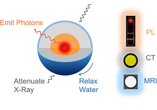
Almutairi and her team recently developed a new nanoparticle with a lanthanide-based core-shell-shell architecture. The nanoparticle emits light for optical imaging, but also relaxes water molecules for MRI and attenuates X-rays for CT simultaneously. 


Three in one: the lanthanide nanoparticles can be used for photoluminescence (PL), computed tomography (CT) and magnetic resonance imaging (MRI) simultaneously.
In a study published in Nano Letters, the researchers tested these nanoparticles in “phantom” tissue — a hydrogel system that mimics living tissue in the laboratory. Not only does the nanoparticle work for each imaging type, it works better than each individual imaging agent on its own.
The team is now working to reduce the size of their new imaging nanoparticle so a patient’s kidneys can clear it more easily from the bloodstream.
“The main point of this study is that we overcame an engineering challenge,” said Sha He, a graduate student in the Jacobs School of Engineering at UC San Diego and first author of the new study. “Now we will tweak the design so we can advance this technology to pre-clinical and clinical testing. Our goal is that one day this nanoparticle, or one like it, will allow a patient to complete his or her imaging all at once, reducing the risk and toxicity associated with separate administration of multiple imaging agents.”

Monday, June 5, 2017

South 8 Technologies wins Clean Tech prize at UC San Diego Entrepreneur Challenge

A new battery technology that will allow electric vehicles to travel farther on a single charge — with significant improvement in safety and cold weather performance — received the Clean Tech top prize of $60,000 in the 10th annual UC San Diego Entrepreneur Challenge pitch competition on May 30, 2017.

The winning technology was developed by a team of engineers and business students at UC San Diego who have founded a new company, called South 8 Technologies, in order to push this technology to market. "We're very grateful for this award and visibility. It's a confidence booster to know that we're developing the right tech at the right time," said UC San Diego materials science and engineering Ph.D. alumnus and South 8 Technologies founder Cyrus Rustomji.

South 8 Technologies was one of six finalist teams that competed in this year's Entrepreneur Challenge. The competition consisted of three tracks: High Tech, Life Tech and Clean Tech. Teams from each track pitched their business plans to local entrepreneurs and professionals with the aim of getting funds to help turn their startups into successful businesses.

The South 8 team developed a technology that enables lithium batteries to run at record low temperatures, down to -60 degrees Celsius (-76 degrees Fahrenheit). In comparison, today's lithium batteries have a low temperature limit of -20 degrees Celsius (-4 degrees Fahrenheit). "One of the big issues with electric vehicles is proper thermal management of the battery pack in cold winter months because batteries do not perform well at low temperature, which can decrease mileage. Our batteries can circumvent this issue," said Jungwoo Lee, a nanoengineering Ph.D. student at UC San Diego and member of South 8 Technologies.

The ability to operate at low temperature is also useful for extreme environment applications such as heavy-duty automotive engine cold-start, high atmosphere WiFi drones or weather balloons, satellites and aerospace applications. The technology can even be extended to ultra-low temperature applications, such as batteries to power spacecraft for interplanetary exploration.

Researchers achieved this exceptional low temperature performance by replacing the conventional liquid electrolyte in lithium batteries with what's called a liquefied gas electrolyte — gas that's stored under mild pressures in the liquid state. "Our liquefied gas electrolytes are conceptually similar to a propane tank for your barbecue grill. Propane is a gas at room temperature and pressure but may be liquefied when stored in a tank under its own vapor pressure," Rustomji said.

"Most work is currently being done in exploring liquid electrolytes and many battery researchers are exploring solid state electrolytes as another alternative. But we're going in the completely opposite direction by exploring gas-based electrolytes."

The technology offers other advantages as well. Liquefied gas electrolytes can potentially increase the energy density of lithium batteries by 50 to 100 percent, meaning that electric vehicles travel much farther on a single charge. These electrolytes also alleviate a problem called thermal runaway, which is when a battery's internal temperature gets hot enough to set off a dangerous chain of chemical reactions that in turn heat up the battery even more. "Our liquefied gas electrolyte offers a unique method of mitigating this thermal runaway which makes the battery safer," Rustomji said.

Rustomji came up with the idea to use liquefied gas electrolytes as a graduate student in the Sustainable Power and Energy Center (SPEC) at UC San Diego under professor Shirley Meng. After graduating with his Ph.D. in 2015, Rustomji continued on as a postdoc to further improve the novel battery chemistry and later joined with Lee and two other engineers in Meng’s lab who soon after became part of the core South 8 Technologies team.

"I couldn't be more grateful for this team," Rustomji said. "They often credit me with inventing this technology, but it's always been a team effort from the start. I owe the early success of South 8 Technologies to our talented engineers and advisor Professor Meng, who have been incredibly supportive, and to the MBA students and business advisor on our team who have been helpful in shaping our business focus."

Lee also noted that part of the team's success stems from a pilot program launched by the UC San Diego Institute for the Global Entrepreneur. Lee and several other members of South 8 Technologies are part of the first class of this pilot program, which teams Jacobs School of Engineering graduate students with MBA students in the Rady School of Management and teaches these students how to develop a business plan. 

"The classes taught us how to better pitch our tech to business people," Lee said. "As engineers, we often describe a technological advance as something that's X percentage better than Y. But to succeed in the business world, we need to describe our tech as something that results in greater value to the customer — something that is worth replacing the existing product."

One of the big selling points made by the South 8 team is that their new electrolyte can be seamlessly integrated into existing battery manufacturing processes. "I think what caught the judges' attention at the Entrepreneur Challenge is that we've developed an inexpensive, drop-in replacement electrolyte which is compatible with conventional batteries being made today. There's no need to redesign an entire manufacturing supply line. I think this is where so many other battery startups have failed," Rustomji said.

Tuesday, September 13, 2016

These fish weren't just made for swimming

There's something fishy going on in the Department of NanoEngineering at UC San Diego. Last summer, 3D-printed microfish made a big splash in the news. Now, researchers have created "nanofish" -- tiny metallic fish 100 times smaller than a grain of sand -- that could be used to deliver drugs to particular places in the body.

The nanofish are made of gold and magnetic nickel segments, which enable the fish to move and be guided by an external magnet.

"We believe they could be useful for medicine delivery, non-invasive surgery and single cell manipulation," said Jinxing Li, a PhD student in the lab of nanoengineering professor Joseph Wang who led the research.

See a video of the nanofish featured in New Scientist:


Monday, September 12, 2016

UC San Diego nanoengineer makes Popular Science’s ‘Brilliant 10’ list


Popular Science magazine has named Liangfang Zhang, a nanoengineering professor at the University of California San Diego, in its 15th annual “Brilliant 10” list, a lineup of “the 10 most innovative young minds in science and engineering.” Zhang was recognized for his revolutionary work in the field of nanomedicine, which focuses on nanomaterials for medical applications.

Zhang invented a way to make nanoparticles perform therapeutic tasks in the body, like treat injuries and deliver drugs to specific sites, without being rejected by the immune system. By coating nanoparticles with natural cell membranes from the body, like red blood cell membranes and platelet cell membranes, researchers can disguise nanoparticles as the body’s own cells.

“We’re essentially camouflaging nanoparticles to look and act like they belong in the body. We want to mimic the natural interaction of the body’s cells with the immune system in order to make new biomimetic nanoparticles that can safely function and survive in the body for long periods of time,” Zhang said.

Zhang’s cell membrane coating technology made its debut in a 2011 study, in which his team showed a new way to disguise nanoparticles as red blood cells. The method involved collecting the membranes from red blood cells and wrapping them around polymeric nanoparticles. This work was an important first step toward a nanodevice—for applications like drug delivery—that could circulate in the body for extended periods without being attacked by the immune system. Since red blood cells live in the body for up to 120 days, Zhang figured they would be good models and resources for making long-circulation drug delivery nanodevices.

In the 2011 study, Zhang’s team showed that nanoparticles coated with red blood cell membranes circulated in the bodies of mice for up to two days. This was an improvement over other nanoparticle systems developed for drug delivery—these are coated with a synthetic material made to temporarily suppress immune recognition and circulate in the body for just a few hours. Zhang explained that a major concern of the synthetic coating is that, after repeated use, it will eventually trigger an immune response and in the long run, these types of drug delivery systems could be drastically less effective.

Using the body’s own red blood cells marked a major breakthrough in the field of drug delivery research. Trying to mimic the most important properties of a red blood cell in a synthetic coating requires an in-depth biological understanding of how all the proteins and lipids function on the surface of a cell. It also poses what many researchers consider an insurmountable technical challenge—recreating that same cell surface precisely in the lab. But Zhang’s approach was to just take the whole surface membrane from an actual red blood cell.

“We approached this problem using an engineering shortcut and bypassed all of this fundamental biology and these technical challenges,” Zhang said. “We don’t need to fully understand exactly what is going on at the protein level. We can just take the entire cell membrane, coat it onto a nanoparticle surface, and make the nanoparticle look like a red blood cell.”

And this red blood cell disguise offers more than just extended circulation time in the body. Because red blood cells are one of the primary targets of pore-forming toxins, such as those produced by MRSA (methicillin-resistant Staphylococcus aureus), Zhang reasoned that his faux red blood cells could also serve as decoys to lure these toxins away.

Indeed, his team showed that nanoparticles coated with red blood cell membranes were capable of removing MRSA toxins from the bloodstream and as a result, also helped clear up infections caused by MRSA bacteria. This is essentially a new way to combat hard to treat bacterial infections—without the use of antibiotics, Zhang said.

Zhang describes himself as a chemical engineer with biomedical interests. He earned his bachelor’s and master’s degrees in chemical engineering at Tsinghua University in China, then earned his PhD in chemical and biomolecular engineering at the University of Illinois at Urbana Champaign, where his research focused on the fundamental science of cell membrane proteins and lipids. Afterwards, he pursued his postdoc at MIT, where his research on lipid-polymer nanoparticles for drug delivery was more applications focused.

“It felt like a natural fit to integrate my training in fundamental chemical engineering principles with biomedical applications,” Zhang said. “I see many parallels between the two fields. I think that studying the flow of nanoparticles through a blood vessel is similar, albeit more complex, to studying the flow of materials through a pipeline. They both involve knowledge of fluid dynamics, thermodynamics, and diffusion laws. Once you know all these fundamental principles, the work makes more sense.”

In 2008, Zhang became an assistant professor at UC San Diego. He was one of the first faculty recruited for the university’s newly formed Department of NanoEngineering. Shortly after, his idea to combine natural cell membranes with synthetic nanoparticles for drug delivery and other biomedical applications was born.

Over the past five years, Zhang and his lab have taken their cell membrane coating technology to new heights. They’ve disguised nanoparticles as human platelets, which have a natural affinity for binding to damaged blood vessels and certain pathogens in the body, like MRSA bacteria. Because of this affinity, platelet-mimicking nanoparticles could be used for targeted drug delivery.

Zhang’s team conducted several experiments. In one, they packed platelet membrane coated nanoparticles with a drug used to heal damaged arteries and administered them to wounded rats; in another experiment, they packed the nanoparticles with antibiotics and administered them to mice infected with MRSA bacteria. In both cases, the drugs were delivered primarily to the affected areas. “That shows the power and the promise of targeted delivery,” Zhang said.

Zhang’s team has also made disguises out of the membranes of beta cells, which are insulin-producing cells in the pancreas. They coated a nanofiber with beta cell membranes to create a pancreas-like microenvironment that encouraged beta cells to congregate, grow and produce more insulin. This work could lead to new treatments for patients with diabetes. “This is another example of mimicking natural interactions in the body to create more effective therapies,” Zhang said.

But researchers are not stopping there. Next on their list is using cell membrane coating technology to develop new systems for combating cancer tumors. Zhang is also working with several biopharmaceutical companies in San Diego to manufacture the red blood cell coated nanoparticles at large scales and get them into clinical trials.

Zhang and the rest of the “Brilliant 10” are featured in the September/October issues of Popular Science magazine and online at http://www.popsci.com/brilliant-10-2016.

Friday, August 26, 2016

Jacobs School alumnus named to MIT Tech Review 35 list

Wei Gao, who earned his PhD in the research group of nanoengineering professor Joe Wang here at the Jacobs School, has been named to the MIT Tech Review's list of Top 35 Innovators Under 35, better known as the TR 35.

Gao, 31, is currently a postdoctoral fellow at UC Berkeley. There he continues to work on technologies similar to those developed by the Center for Wearable Sensors, led by Wang here at the Jacobs School.

In January 2016, he was first author on a Nature paper titled "Fully integrated wearable sensor arrays for multiplexed in situ perspiration analysis." The study received broad media coverage, including in The New York Times, The Wall Street Journal, Scientific American, IEEE Spectrum, Wired and many other outlets.

Essentially, Gao and colleagues built a sweatband that combines sensors with electronic processors and a Bluetooth transmitter on a flexible printed circuit board. The device wirelessly transmits data about what's in your sweat to an app on your cell phone.

Gao told MIT Tech Review:

“I grew up in a small village in Xuzhou, China. When I was a child I saw a lot of people around me dying of different diseases. Many people don’t realize there’s a problem until it’s too late. I thought, in the future I should design a wearable electronic device to monitor health and tell us what’s going on and what’s going wrong before it gets bad.

“Our body is generating data all the time. There are so many wearable devices now—the Apple watch, the Fitbit—but they mainly track physical activities or vital signs. They can’t provide information at the molecular level.

“It came into my mind: what about sweat?”

Thursday, August 11, 2016

Caleb Christianson, a JPL intern and NanoEngineering Ph.D. student, takes over the Jacobs School's Instagram for a day

Summer interns from UC San Diego. These students represent multiple majores, including computer science and engineering, chemical engineering, electrical engineering, environmental engineering, nanoengineering, and Earth sciences.

From left to right: Diana Alsindy, Christian Gutierrez, Hannah Munguia, David Ruble, Neil Chandiramani, Nicholas Lopez, Annie Chen, Nolan Fewell, Kyle Mirzakhanian, Rahul Palamuttam, Matt Epperson, Leon Cheung, and Caleb Christianson. 

Nanoengineering Ph.D. student Caleb Christianson took over the Jacobs School Instagram account (@ucsandiegoengineering) yesterday, August 10, to give us a preview of what it's like to be a NASA Jet Propulsion Laboratory intern designing and robot that can hover above asteroids! In case you missed it, here's a recap:


A video posted by UC San Diego Engineering (@ucsandiegoengineering) on

A photo posted by UC San Diego Engineering (@ucsandiegoengineering) on


Here is more about the project Caleb worked on this summer, via JPL's newsletter:

Glide, don't land, to study an asteroid
There's intense interest in studying asteroids, but the challenge is getting near them. There isn't enough gravity or atmosphere to allow a spacecraft to touch down without ricocheting off the surface.
But why land when you can glide? JPLer Marco Quadrelli's Electrostatic Glider (E-Glider) was inspired by the idea of dust fountains visible on our moon's surface: when warmed by the sun, these dust particles gain an electrostatic charge. The same principle turns every asteroid or comet's dust into a weak but usable power supply.
Quadrelli's glider would be a low-cost craft attached to foil-like streamers. These streamers would inflate and lift based on the electrostatic energy around them. The glider could then be steered around an asteroid and perform basic science readings on its composition


Caleb Christianson, a JPL intern and NanoEngineering PhD student, takes over the Jacobs School's Instagram for a day


Nanoengineering Ph.D. student Caleb Christianson took over the Jacobs School Instagram account (@ucsandiegoengineering) yesterday, August 10, to give us a preview of what it's like to be a NASA Jet Propulsion Laboratory intern designing and robot that can hover above asteroids! In case you missed it, here's a recap:


#JPLInternTakeover

A video posted by UC San Diego Engineering (@ucsandiegoengineering) on

UCSD JPL interns represent! #SelfieStyle #JPLInternTakeover

A photo posted by UC San Diego Engineering (@ucsandiegoengineering) on